
Pulmonary emergencies on USMLE Step 2 CK test whether you choose the right diagnostic test and recognize when a patient needs immediate intervention. Try these three questions, then open each explanation.
Question 1: Sudden shortness of breath after hip surgery
A 54-year-old woman develops sudden shortness of breath and right-sided pleuritic chest pain 5 days after a total hip replacement. Pulse is 112/min, respirations are 24/min, blood pressure is 128/80 mm Hg, and oxygen saturation is 89% on room air. The lungs are clear. Her serum creatinine is normal, and she has no history of contrast allergy. Which of the following is the most appropriate next step to confirm the diagnosis?
A. D-dimer level
B. Lower extremity compression ultrasonography
C. CT pulmonary angiography
D. Ventilation-perfusion scan
E. Transthoracic echocardiography
Question 2: An asthma attack with a “normal” PaCO2
A 24-year-old man with asthma has had increasing shortness of breath for 2 days. He has received three nebulized albuterol and ipratropium treatments and intravenous methylprednisolone. He is now drowsy and can speak only one or two words at a time. Breath sounds are barely audible, with almost no wheezing. Arterial blood gas on oxygen shows pH 7.32, PaCO2 47 mm Hg and PaO2 62 mm Hg. Which of the following is the most appropriate next step?
A. Discharge home with an oral steroid taper
B. Add montelukast
C. Add inhaled salmeterol
D. Chest physiotherapy
E. Endotracheal intubation and mechanical ventilation
Question 3: Hypotension and absent breath sounds after a car crash
A 30-year-old man is brought to the emergency department after a high-speed motor vehicle collision. He is in severe respiratory distress. Blood pressure is 76/40 mm Hg and pulse is 136/min. Breath sounds are absent on the left side, the left chest is hyperresonant to percussion, the trachea is shifted to the right, and the neck veins are distended. Which of the following is the most appropriate next step?
A. Chest x-ray
B. Needle decompression or finger thoracostomy of the left chest
C. CT scan of the chest
D. Pericardiocentesis
E. Intravenous fluids and observation
Key takeaways
- High-probability PE with normal kidneys: CT pulmonary angiography (D-dimer only for low probability).
- Asthma attack with normal or rising PaCO2, drowsiness or a silent chest = impending respiratory failure: intubate.
- Tension pneumothorax is a clinical diagnosis: decompress immediately, then place a chest tube.
More USMLE practice questions
- All USMLE Step 2 CK questions
- Newborn care, stroke airway and myelomeningocele
- Psychiatry: panic disorder and agoraphobia
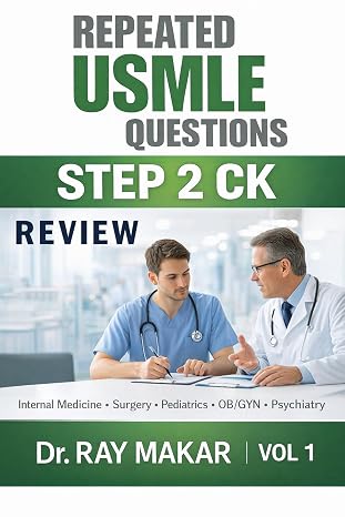
Want 100+ more clinical questions like these?
Get Repeated USMLE Questions Step 2 CK Review Vol 1 by Dr. Ray Makar
As an Amazon Associate, I earn from qualifying purchases.
Written and reviewed by Dr. Ray Makar, MD, dermatologist with 20 years of clinical practice. Last reviewed: October 2026. For exam preparation only, not medical advice.
FREE USMLE PDFs
Get 80 free practice questions
Four free sampler PDFs by Dr. Ray Makar: Step 1 (Pathology & Pharmacology) and Step 2 CK (Medicine, Surgery & Pediatrics), in both Review style and USMLE style, with answers and explanations. Also useful for NEET-PG, INI-CET and FMGE.
Download the free PDFs See the full books →