
Neurology emergencies on USMLE Step 2 CK test time-sensitive decisions: when to give thrombolytics, which test to order after a normal CT, and when to treat before the diagnosis is confirmed. Try these three questions.
Question 1: Sudden weakness and trouble speaking
A 67-year-old man is brought to the emergency department 2 hours after the sudden onset of right arm and leg weakness and difficulty speaking. Blood pressure is 168/95 mm Hg. Fingerstick glucose is 110 mg/dL. Noncontrast CT of the head shows no hemorrhage. He takes no anticoagulants and has no recent surgery, bleeding or prior intracranial hemorrhage. Which of the following is the most appropriate next step in treatment?
A. Aspirin 325 mg and observation
B. Intravenous heparin infusion
C. Lower the blood pressure below 140/90 mm Hg before any other treatment
D. MRI of the brain before treatment
E. Intravenous thrombolytic therapy (alteplase or tenecteplase)
Question 2: The “worst headache of my life” with a normal CT
A 45-year-old woman comes to the emergency department because of a sudden, severe headache that began 10 hours ago while she was exercising. She describes it as the worst headache of her life and reached maximal intensity within a minute. She has neck stiffness. Neurologic examination is otherwise normal. Noncontrast CT of the head shows no abnormalities. Which of the following is the most appropriate next step?
A. Discharge with analgesics
B. Lumbar puncture
C. Electroencephalography
D. Subcutaneous sumatriptan
E. Repeat CT of the head in 1 week
Question 3: New headache and jaw pain while chewing in an older woman
A 72-year-old woman has had a new headache over her right temple for 3 weeks. Her scalp is tender when she combs her hair, and her jaw aches while chewing. She also has morning stiffness in her shoulders and hips. Visual acuity is normal. The right temporal artery is tender and has a decreased pulse. ESR is 92 mm/h. Which of the following is the most appropriate next step?
A. Temporal artery biopsy, with treatment only after the result
B. MRI of the brain
C. Naproxen
D. High-dose oral prednisone, started now
E. Low-dose prednisone (10–15 mg daily)
Key takeaways
- Ischemic stroke within 4.5 hours, no hemorrhage, BP below 185/110: IV thrombolysis; check for large vessel occlusion for thrombectomy.
- Thunderclap headache with negative CT after 6 hours: lumbar puncture for xanthochromia.
- Giant cell arteritis: start high-dose steroids immediately, then confirm with biopsy.
More USMLE practice questions
- All USMLE Step 2 CK questions
- Newborn care, stroke airway and myelomeningocele
- Psychiatry: panic disorder and agoraphobia
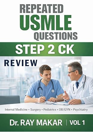
Want 100+ more clinical questions like these?
Get Repeated USMLE Questions Step 2 CK Review Vol 1 by Dr. Ray Makar
As an Amazon Associate, I earn from qualifying purchases.
Written and reviewed by Dr. Ray Makar, MD, dermatologist with 20 years of clinical practice. Last reviewed: October 2026. For exam preparation only, not medical advice.
FREE USMLE PDFs
Get 80 free practice questions
Four free sampler PDFs by Dr. Ray Makar: Step 1 (Pathology & Pharmacology) and Step 2 CK (Medicine, Surgery & Pediatrics), in both Review style and USMLE style, with answers and explanations. Also useful for NEET-PG, INI-CET and FMGE.
Download the free PDFs See the full books →